loading

Expert Pilonidal Sinus Care in Delhi

pilonidal sinus surgery

Treatment for deep vein thrombosis (DVT) aims to prevent the clot from growing larger, reduce the risk of complications such as pulmonary embolism (PE), and alleviate symptoms. Here’s an overview of the treatment options and strategies commonly used for managing DVT:
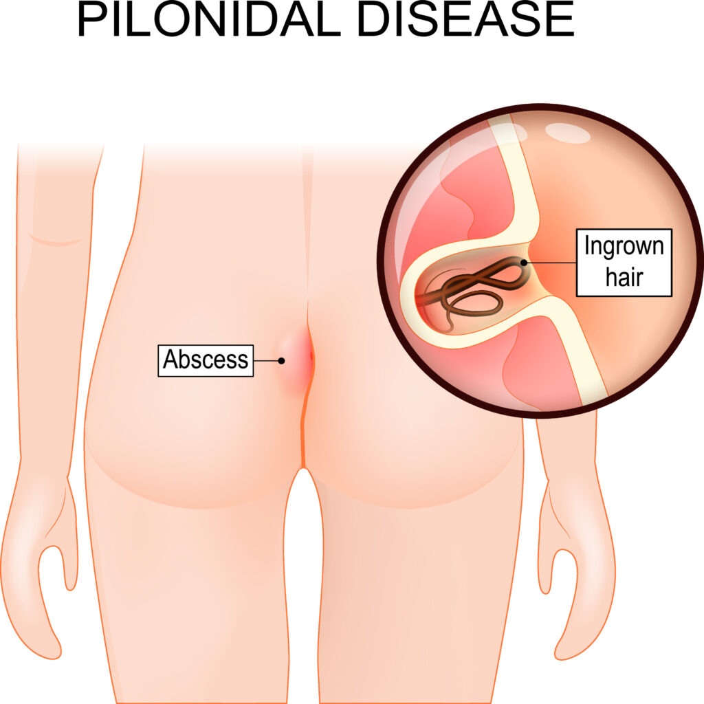
pilonidal sinus surgery

. Anticoagulant Medications

  • Heparin: Often used initially to rapidly thin the blood and prevent further clotting. It is administered intravenously in the hospital or subcutaneously at home.
  • Warfarin (Coumadin): A vitamin K antagonist taken orally. It is started alongside heparin and continued for several months to prevent clot recurrence. Regular blood tests (INR monitoring) are necessary to adjust the dose.
  • Direct Oral Anticoagulants (DOACs): These newer medications, such as rivaroxaban (Xarelto), apixaban (Eliquis), edoxaban (Savaysa), and dabigatran (Pradaxa), are taken orally and work directly on specific clotting factors in the blood. They may be used instead of warfarin in some cases.
  1. Thrombolytic Therapy
  • Thrombolytics (Clot-busting Medications): Used in severe cases of DVT or when there is a high risk of complications like limb ischemia. These medications are administered through a catheter directly into the clot to dissolve it quickly. This treatment is more aggressive and carries a higher risk of bleeding complications.
  1. Compression Stockings
  • Compression Therapy: Graduated compression stockings or wraps are worn on the affected leg to help reduce swelling and improve blood flow. They are typically worn during the day and removed at night.
  1. Inferior Vena Cava (IVC) Filter
  • IVC Filter: A small device inserted into the inferior vena cava (large vein in the abdomen) to trap blood clots that may break loose from leg veins and prevent them from traveling to the lungs. It is considered when anticoagulation is contraindicated or ineffective, or when there is a recurrence of PE despite adequate anticoagulation.
  1. Catheter-directed Thrombolysis and Thrombectomy
  • Catheter-directed Thrombolysis: Similar to thrombolytic therapy, but delivered through a catheter directly into the clot for more localized treatment.
  • Thrombectomy: Surgical removal of the clot, which is reserved for severe cases of DVT or when other treatments are not effective.
  1. Lifestyle and Home Care
  • Activity Modification: Avoiding prolonged sitting or standing. Regular movement and exercises that promote blood flow in the legs.
  • Elevation: Keeping the affected leg elevated when resting to reduce swelling.
  • Hydration: Staying well-hydrated to maintain healthy blood circulation.
  • Smoking Cessation: Quitting smoking to reduce the risk of clot formation and improve overall vascular health.
  1. Follow-Up and Monitoring
  • Regular Follow-Up: Monitoring with healthcare providers to assess response to treatment, manage medication adjustments, and screen for complications.
  • Imaging Studies: Follow-up ultrasound or venography to evaluate clot resolution and check for recurrent DVT.

Complications and Considerations

  • Bleeding: Anticoagulant therapy increases the risk of bleeding, which can be serious. Close monitoring and adjustments in medication doses are necessary.
  • Post-thrombotic Syndrome: Chronic leg pain, swelling, and skin changes due to damage to the veins and valves after DVT.
  • Pulmonary Embolism: Risk of PE remains until the clot is adequately treated and resolved.
  • Recurrence: Even with treatment, DVT can recur, especially if risk factors persist.

Conclusion

Effective management of deep vein thrombosis requires a tailored approach based on the severity of the clot, presence of symptoms, and individual health factors. Early diagnosis, prompt initiation of anticoagulant therapy, and appropriate follow-up are crucial for reducing complications and improving outcomes in patients with DVT. Lifestyle modifications and adherence to treatment recommendations play key roles in preventing recurrence and promoting overall vascular health.

3.5

pilonidal sinus surgery

ChatGPT

Pilonidal Sinus Surgery

Pilonidal sinus surgery is performed to treat a pilonidal sinus, a small cyst or abscess that occurs in the cleft at the top of the buttocks. This condition often involves infection and can cause significant discomfort. Here’s an overview of the surgical treatment for a pilonidal sinus, including indications, types of surgery, the procedure, and postoperative care:

Indications

Surgical intervention is indicated for:

  • Recurrent or chronic pilonidal sinus infections.
  • Abscess formation that doesn’t respond to antibiotics and drainage.
  • Persistent pain or drainage from the sinus.
  • Failure of conservative treatments.

Types of Pilonidal Sinus Surgery

There are several surgical options for treating a pilonidal sinus, ranging from minimally invasive procedures to more extensive surgeries:

  1. Incision and Drainage (I&D):
    • Indication: Used for acute abscesses.
    • Procedure: The abscess is incised and drained. The cavity is then packed with gauze to allow for healing from the inside out.
    • Postoperative Care: Regular dressing changes until the wound heals.
  2. Excision and Primary Closure:
    • Indication: Chronic or recurrent pilonidal disease.
    • Procedure: The sinus and surrounding tissue are excised, and the wound is closed with sutures.
    • Advantages: Faster healing compared to open wound techniques.
    • Disadvantages: Higher risk of wound infection and recurrence.
  3. Excision with Secondary Intention Healing:
    • Indication: Preferred for minimizing recurrence.
    • Procedure: The sinus and surrounding tissue are excised, but the wound is left open to heal naturally.
    • Advantages: Lower recurrence rates.
    • Disadvantages: Longer healing time and more intensive wound care.
  4. Flap Procedures:
    • Indication: Recurrent or complex pilonidal disease.
    • Procedure: After excision of the sinus, a flap of healthy tissue (such as the Limberg or Karydakis flap) is rotated and sutured to cover the defect.
    • Advantages: Lower recurrence rates and quicker healing.
    • Disadvantages: More complex surgery requiring expertise.
  5. Minimally Invasive Techniques:
    • Laser Surgery: Laser is used to ablate the sinus tract.
    • Endoscopic Pilonidal Sinus Treatment (EPSiT): Uses a small endoscope to visualize and remove the sinus tract.

Procedure Steps

  1. Preoperative Preparation:
    • Medical evaluation and history taking.
    • Imaging studies if needed to map the extent of the sinus.
    • Instructions on fasting and medication adjustments if required.
  2. Anesthesia:
    • Local, regional, or general anesthesia depending on the extent of the surgery and patient preference.
  3. Surgical Procedure:
    • Positioning: Patient is positioned prone (lying on the stomach).
    • Incision: The surgeon makes an incision over the affected area.
    • Excision: The sinus tract and any infected tissue are removed.
    • Closure: Depending on the chosen technique, the wound may be closed with sutures, left open, or covered with a flap.
  4. Postoperative Care:
    • Wound Care: Instructions on keeping the wound clean and dry. Regular dressing changes for open wounds.
    • Pain Management: Prescribed pain relief medications.
    • Activity Restrictions: Limited physical activity to avoid strain on the surgical site.
    • Follow-Up: Regular follow-up visits to monitor healing and detect any complications early.

Risks and Complications

  • Infection: Risk of wound infection post-surgery.
  • Recurrence: Pilonidal sinuses can recur even after surgery, particularly if proper wound care is not maintained.
  • Scarring: There will be some scarring, the extent of which depends on the surgical technique used.
  • Delayed Healing: Particularly with open wound techniques.

Conclusion

Surgical treatment for pilonidal sinus aims to remove the infected tissue and prevent recurrence. The choice of surgical technique depends on the individual case, with considerations for the patient’s health, the extent of the disease, and the surgeon’s expertise. Proper postoperative care, including wound management and follow-up, is crucial for a successful outcome and to minimize the risk of complications.