loading

Retinal Detachment Eye Care Treatment in Delhi

Retinal Detachment Surgery

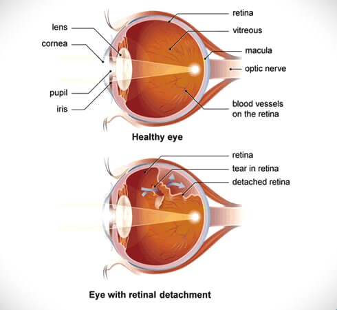
Retinal detachment surgery is a procedure designed to reattach the retina to its correct position at the back of the eye. The retina is a thin layer of tissue lining the back of the eye, essential for vision as it captures light and sends visual signals to the brain. Retinal detachment happens when this layer is pulled away from its normal position, resulting in vision loss.

retinal detachment
Common Types of Retinal Detachment Surgery
  • Pneumatic Retinopexy:
    • Procedure: A gas bubble is injected into the vitreous cavity of the eye. The patient then positions their head so that the bubble floats to the site of the retinal tear, pushing the retina back against the wall of the eye. Laser or cryopexy (freezing) treatment is then used to seal the tear.
    • Indications: Suitable for certain types of retinal detachment, typically those with small, uncomplicated tears located in the upper part of the retina.
    • Recovery: Requires the patient to maintain a specific head position for several days to keep the bubble in the correct position.
  • Scleral Buckling:
    • Procedure: A flexible band (scleral buckle) is placed around the eye to counteract the force causing the retinal detachment. The buckle indents the wall of the eye, relieving some of the pull on the retina. Laser or cryopexy treatment is also used to seal the tear.
    • Indications: Effective for a wide range of retinal detachments, particularly those caused by more extensive retinal tears.
    • Recovery: Post-operative discomfort is common, and vision may take a while to improve. Activity restrictions are typically required.
  • Vitrectomy:
    • Procedure: The vitreous gel pulling on the retina is removed and replaced with a gas bubble, silicone oil, or saline solution. The retina is then treated with laser or cryopexy to seal the tear.
    • Indications: Often used for more complex retinal detachments, including those involving significant scar tissue or bleeding in the vitreous.
    • Recovery: Depending on the type of replacement used, specific head positioning may be necessary. Recovery time varies, and vision improvement can take weeks to months.
  • Laser Surgery (Photocoagulation):
    • Procedure: A laser is used to create small burns around the retinal tear, creating scar tissue that helps seal the tear and prevent fluid from passing through.
    • Indications: Suitable for small retinal tears or holes that can lead to detachment.
    • Recovery: Typically has a quicker recovery time compared to other procedures but is only suitable for certain cases.
Recovery and Post-Operative Care
  • Monitoring: Regular follow-up visits are crucial to monitor the healing process and ensure the retina remains attached.
  • Activity Restrictions: Patients may need to avoid activities that could increase eye pressure or cause trauma to the eye.
  • Vision Improvement: Vision recovery varies; it can take weeks to months, and some vision loss may be permanent, depending on the severity of the detachment and the timeliness of the treatment.
Potential Complications
  • Infection
  • Bleeding
  • Increased intraocular pressure
  • Cataract formation
  • Recurrence of retinal detachment

Timely treatment of retinal detachment is critical to prevent permanent vision loss. If you experience symptoms such as sudden flashes of light, floaters, or a shadow over your vision, seek immediate medical attention.